Skin cancer examination and diagnostics

How is skin cancer evaluated?
Your primary care physician can perform the initial evaluation, but you may also be referred to a skin specialist, called a dermatologist.
Doctors first evaluate skin changes based on your medical history, including your description of symptoms and how the lesion has developed over time. The doctor will also examine the skin lesion using a dermatoscope, a handheld device that provides magnification and illumination to allow detailed visualization of skin structures.
If the doctor determines that further evaluation is needed, a skin sample (biopsy) may be taken under local anesthesia. This may be performed as a small punch biopsy or by removing the entire suspicious lesion (excisional biopsy). In some cases, the doctor may also examine nearby lymph nodes to check for enlargement.
You will usually receive immediate feedback from the clinical examination and dermoscopy, as well as from the lymph node examination. However, a biopsy must be analyzed in a laboratory, and results are typically available within a few weeks.
What does the examination mean for you as a patient?
Examination with a dermatoscope and evaluation of lymph nodes are painless and provide immediate information. If a biopsy is performed, local anesthesia is typically used. The procedure may cause mild discomfort and can leave a small scar. Waiting for biopsy results may understandably cause concern for some patients.

Examination through a dermascope.
New imaging technologies
Research is ongoing into noninvasive imaging techniques that allow the skin to be examined using optical technologies without the need for a biopsy. These methods can help define the extent of a tumor, assess whether a biopsy is necessary, and in some cases assist in making a diagnosis more quickly.
In Denmark, these technologies are currently used primarily in research settings, for example to monitor treatment response and to identify imaging features that correlate with biopsy findings.
Interpretation of these images requires specialized expertise. Artificial intelligence (AI) is also being developed to assist with image analysis and help support physicians in diagnosis and clinical decision-making.
Advantages of noninvasive imaging include:
- Faster diagnosis and the potential for treatment during the same visit
- Fewer biopsies and less discomfort for patients
- More precise definition of tumor boundaries before treatment
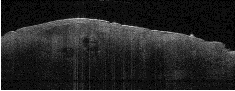
Optic imaging during a scan for skin cancer
How is skin cancer diagnosed?
The diagnosis of skin cancer is based primarily on clinical evaluation and confirmation with a biopsy. New imaging technologies can provide important additional information and may help reduce waiting times and improve patient comfort in the future.
A diagnosis of skin cancer is confirmed when a skin biopsy examined in a laboratory shows cancer cells. Laboratory analysis determines whether cancer is present, the specific type of skin cancer, and, in some cases, important tumor characteristics. This information is essential for selecting the most appropriate treatment.