New research could pave the way for improved diagnostics and treatment choices for actinic keratoses

Today, doctors evaluate actinic keratoses (AK) using a classification system that divides lesions into three grades based on the thickness of the skin changes. However, this method has its limitations, as there is no correlation between what is seen when examining tissue samples under a microscope. Additionally, the assessment can be subjective and vary from one doctor to another.
To improve the clinical evaluation of AK, researchers have explored advanced non-invasive technology called 'dynamic optical coherence tomography' (D-OCT). This technique allows for ‘looking’ beneath the skin and measuring the structure of blood vessels, which could prove to be a game-changer. The studies reveal that the complexity of blood vessels changes significantly as AK lesions thicken and in comparison to nearby normal skin.
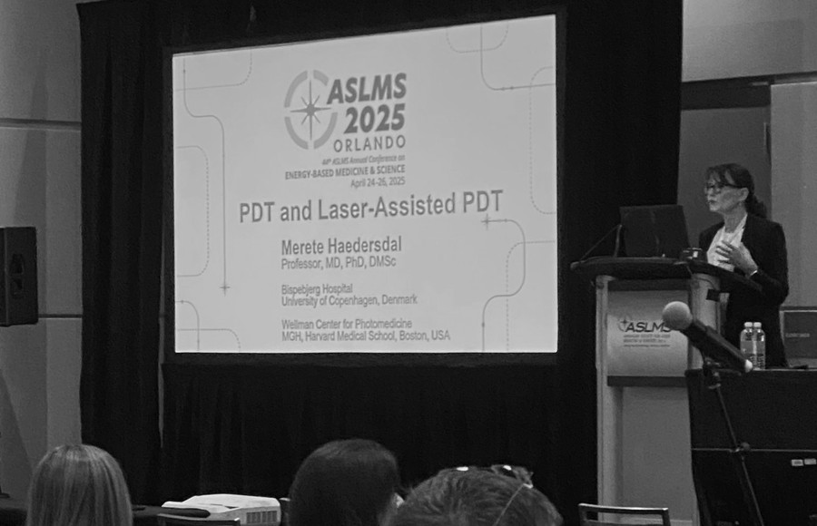
The research project was carried out by physician and PhD student Gabriela Fredman, along with supervisors from Bispebjerg Hospital, Professor Merete Haedersdal, and from DTU, postdoc Gavrielle Untracht. The studies examined the characteristics of small blood vessels across the three AK grades and in sun-damaged skin in 47 test subjects. The results, published in two studies, show how manual analyses of D-OCT images can provide better insights and potentially help develop automated calculation models, making the assessments even more precise.
It is hoped that D-OCT could become a valuable clinical tool for faster diagnosis, more precise monitoring of treatment effects, and potentially improving the choice of treatment for patients with actinic keratoses.


